Primary Antibody Deficiency Panel
Summary and Pricing 
Test Method
Exome Sequencing with CNV Detection| Test Code | Test Copy Genes | Panel CPT Code | Gene CPT Codes Copy CPT Code | Base Price | |
|---|---|---|---|---|---|
| 2687 | Genes x (46) | 81479 | 81321(x1), 81323(x1), 81403(x1), 81404(x2), 81406(x1), 81408(x1), 81479(x85) | $990 | Order Options and Pricing |
Pricing Comments
We are happy to accommodate requests for testing single genes in this panel or a subset of these genes. The price will remain the list price. If desired, free reflex testing to remaining genes on panel is available. Alternatively, a single gene or subset of genes can also be ordered via our Custom Panel tool.
An additional 25% charge will be applied to STAT orders. STAT orders are prioritized throughout the testing process.
Click here for costs to reflex to whole PGxome (if original test is on PGxome Sequencing platform).
Click here for costs to reflex to whole PGnome (if original test is on PGnome Sequencing platform).
Turnaround Time
3 weeks on average for standard orders or 2 weeks on average for STAT orders.
Please note: Once the testing process begins, an Estimated Report Date (ERD) range will be displayed in the portal. This is the most accurate prediction of when your report will be complete and may differ from the average TAT published on our website. About 85% of our tests will be reported within or before the ERD range. We will notify you of significant delays or holds which will impact the ERD. Learn more about turnaround times here.
Targeted Testing
For ordering sequencing of targeted known variants, go to our Targeted Variants page.
Clinical Features and Genetics 
Clinical Features
Primary antibody deficiencies encompass a heterogeneous group of B-cell immunodeficiencies marked by reduced or absent antibody production. These disorders are highlighted by increased susceptibility to infection. The three categories of antibody deficiencies are common variable immunodeficiency (CVID), hyper IgM syndromes (HIGM) and agammaglobulinemia (AGG) (Bousfiha et al. 2018. PubMed ID: 29226301; Picard et al. 2018. PubMed ID: 29226302).
CVID is the most common primary immune deficiency affecting one in ~25,000 births. CVID is an immunodeficiency disorder hallmarked by impaired B cell differentiation and antibody production, namely IgG and IgA. CVID is primarily diagnosed via exclusion of other primary immune disorders. However, recurrent bacterial infections after two years of age, poor response of vaccines, and decreased IgG and IgA are indicative of CVID (Conley et al. 1999. PubMed ID: 10600329). About half of CVID patients also present with decreased serum IgM. CVID is a heterogeneous disorder affecting multiple organ systems and with symptoms ranging from autoimmune disease, chronic lung disease, bronchiectasis, gastrointestinal disease, malabsorption, granulomatous disease, liver disease, and/or lymphoma (Resnick et al. 2012. PubMed ID: 22180439; Conley et al. 2009. PubMed ID: 19302039). Due to the highly variable clinical presentation of CVID, median diagnostic delay is 4.1 years with 20% of patients being diagnosed before 20 years of age. The majority of patients with CVID are diagnosed between the ages of 20-45 years (Gathmann et al. 2014 PubMed ID: 24582312).
HIGM is due to impaired immunoglobulin class switch recombination (CSR) resulting in an increase in IgM and a decrease in IgG, IgA and IgE antibody levels. This humoral immunodeficiency leads to recurrent sinopulmonary infections, chronic diarrhea due to Cryptosporidium infection, and neutropenia presenting within the first or second decade of life. In severe cases, patients may also present with, sclerosing cholangitis, chronic arthritis, thrombocytopenia, hemolytic anemia, hypothyroidism, kidney disease and failure to thrive (Davies and Thrasher. 2010. PubMed ID: 20180797; Etzioni and Ochs. 2004. PubMed ID: 15319456; Leven et al. 2016. PubMed ID: 27189378).
Patients with agammaglobulinemia present with recurrent bacterial infections including Haemophilus influenza, Streptococcus pneumoniae, and staphylococci due to loss of antibody mediated immune responses. Common sites of infection include skin, respiratory tract, gastrointestinal tract, and joints. Severe infections can be life threatening due to infections spreading leading to empyema, meningitis, and sepsis. Infections typically occur after the first few months of life as maternal immunoglobulins are protective (Conley and Howard. 2011. PubMed ID: 20301626). Diagnosis primarily occurs in the first two years of life, although 10% of cases have delayed onset with immunodeficiency not being apparent until after ten years of age.
Genetics
In about 75% of CVID, a molecular diagnosis is not found. This is due to the multifactorial etiology of CVID including both genetic and environmental factors. Autosomal dominant CVIDs occur through pathogenic variants in the TNFRSF13B, IRF2BP2, NFKB1, NFKB2, VAV1, TNFSF12 and IKZF1 genes. Autosomal recessive CVIDs occur through pathogenic variants in the ICOS, IL21, CD81, MS4A1, NFKB2, CR2, LRBA, MOGS, TNFRSF13C, SKIC3/TTC37, or CD19 genes (Bousfiha et al. 2018. PubMed ID: 29226301; Picard et al. 2018. PubMed ID: 29226302). About 20% of CVID cases are due to pathogenic variants in the TNFRSF13B gene with ~90% of these cases being sporadic. Two missense variants, c.310T>C (p.Cys104Arg) and c.542C>A (p.Ala181Glu) are most commonly found and represent about 80% of all TNFRSF13B-related CVID cases (Salzer et al. 2009. PubMed ID: 18981294).
HIGM1 is inherited in an X-linked manner through pathogenic variants in the CD40LG gene. HIGM can also be inherited in an autosomal recessive manner through pathogenic variants in the AICDA, CD40, INO80 or UNG genes. About 75% of cases to date are due to pathogenic variants in the CD40LG gene (Johnson et al. 2013. PubMed ID: 20301576). Other syndromes inherited in an X-linked recessive manner that also exhibit increased IgM levels include agammaglobulinemia (BTK), and lymphoproliferative syndrome (SH2D1A). Related autosomal dominant disorders include primary immunodeficiency (PIK3CD) and ectodermal dysplasia (NFKBIA). Autosomal recessive conditions include ataxia telangiectasia (ATM), Nijmegen breakage syndrome (NBN), and ataxia telangiectasia like disorder (MRE11) (Bousfiha et al. 2018. PubMed ID: 29226301; Picard et al. 2018. PubMed ID: 29226302).
X-linked agammaglobulinemia represents about 85% of AGG cases via pathogenic variants in the BTK gene. To date over 700 different pathogenic variants in the BTK gene have been reported with no single pathogenic variant representing more than 3% of XLA cases (Conley et al. 2005. PubMed ID: 15661032; Lindvall et al. 2005. PubMed ID: 15661031; Väliaho et al. 2006. PubMed ID: 16969761). Autosomal recessive forms of AGG are due to pathogenic variants in the BLNK, CD79A, CD79B, IGHM, IGLL1, MOGS, PIK3R1, TRNT1 and SH2D1A genes. AGG via pathogenic variants in the LRRC8 and TCF3 genes are inherited in an autosomal dominant manner (Bousfiha et al. 2018. PubMed ID: 29226301; Picard et al. 2018. PubMed ID: 29226302).
See individual gene test descriptions for information on molecular biology of gene products and mutation spectra.
Clinical Sensitivity - Sequencing with CNV PGxome
In a study of 50 patients with common variable immunodeficiency, a genetic diagnosis was found in 15 cases. Pathogenic variants were identified in the TNFRSF13B (14%), NFKB1 (10%), STAT3 (6%), CTLA4 (4%), PIK3CD (2%), IZKF1 (2%), LRBA (4%), and STXBP2 (2%) genes (Maffucci et al. 2016. PubMed ID: 27379089).
In a molecular study of 140 patients presenting with hyper IgM syndromes based on recurrent and/or severe infections, elevated IgM, and reduced IgA and IgG levels, sequencing of the CD40LG, CD40, NEMO, AICDA, UNG, ICOS, ICOSL, BTK, and SH2D1 genes was performed. Pathogenic variants were found in 103 of 140 patients with pathogenic variants in the CD40LG, AICDA, UNG, NEMO, and BTK genes accounting for 92%, 4%, 1%, 1%, and 3% of cases respectively (Lee et al. 2005. PubMed ID: 15358621).
X-linked agammaglobulinemia accounts for 90% of cases of agammaglobulinemia in males through pathogenic variants in the BTK gene. Autosomal recessive forms of agammaglobulinemia through pathogenic variants in the IGHM, CD79A, CD79B, or BLNK genes represent 10% of cases in males (Conley and Howard. 2011. PubMed ID: 20301626; Conley et al. 1998. PubMed ID: 9545398; Kanegane et al. 2001. PubMed ID: 11742281).
Testing Strategy
This test is performed using Next-Gen sequencing with additional Sanger sequencing as necessary.
This panel typically provides 99.2% coverage of all coding exons of the genes plus 10 bases of flanking noncoding DNA in all available transcripts along with other non-coding regions in which pathogenic variants have been identified at PreventionGenetics or reported elsewhere. We define coverage as ≥20X NGS reads or Sanger sequencing. PGnome panels typically provide slightly increased coverage over the PGxome equivalent. PGnome sequencing panels have the added benefit of additional analysis and reporting of deep intronic regions (where applicable).
Dependent on the sequencing backbone selected for this testing, discounted reflex testing to any other similar backbone-based test is available (i.e., PGxome panel to whole PGxome; PGnome panel to whole PGnome).
Indications for Test
Candidates for testing include individuals with decreased serum immunoglobulin levels for IgG, IgA and/or IgM and normal B cell levels. In the case of HIGM, individuals typically present with decreased IgG and IgA levels, but elevated IgM (Bonilla et al. 2015. PubMed ID: 26371839). Patients with primary antibody deficiencies generally present with recurrent and/or severe respiratory infections, recurrent otitis media, sinusitis and pneumonia. Respiratory infections involving Streptococcus pneumoniae and Haemophilus influenza are common features. Other clinical findings may include failure to thrive, recurrent fevers, chronic diarrhea, nodal lymphoid hyperplasia in the gut or unexplained hepatosplenomegaly. Autoimmune disease may also be present, but is typically found in adults (Bousfiha et al. 2018. PubMed ID: 29226301; Picard et al. 2018. PubMed ID: 29226302).
Candidates for testing include individuals with decreased serum immunoglobulin levels for IgG, IgA and/or IgM and normal B cell levels. In the case of HIGM, individuals typically present with decreased IgG and IgA levels, but elevated IgM (Bonilla et al. 2015. PubMed ID: 26371839). Patients with primary antibody deficiencies generally present with recurrent and/or severe respiratory infections, recurrent otitis media, sinusitis and pneumonia. Respiratory infections involving Streptococcus pneumoniae and Haemophilus influenza are common features. Other clinical findings may include failure to thrive, recurrent fevers, chronic diarrhea, nodal lymphoid hyperplasia in the gut or unexplained hepatosplenomegaly. Autoimmune disease may also be present, but is typically found in adults (Bousfiha et al. 2018. PubMed ID: 29226301; Picard et al. 2018. PubMed ID: 29226302).
Genes
| Inheritance | Abbreviation |
|---|---|
| Autosomal Dominant | AD |
| Autosomal Recessive | AR |
| X-Linked | XL |
| Mitochondrial | MT |
Diseases
Related Tests
| Name |
|---|
| PGxome® |
| Common Variable Immune Deficiency (CVID) Panel |
| Hyper IgM Syndrome Panel |
Citations 
- 8. Davies and Thrasher. 2010. PubMed ID: 20180797
- 10. Salzer et al. 2009. PubMed ID: 18981294
- Bonilla et al. 2015 PubMed ID: 26371839
- Bousfiha et al. 2018 PubMed ID: 29226301
- Conley et al. 1998. PubMed ID: 9545398
- Conley et al. 1999. PubMed ID: 10600329
- Conley et al. 2005 PubMed ID: 15661032
- Conley et al. 2009. PubMed ID: 19302039
- Etzioni and Ochs. 2004. PubMed ID: 15319456
- Gathmann et al. 2014 PubMed ID: 24582312
- Johnson et al. 2013 PubMed ID: 20301576
- Kanegane et al. 2001. PubMed ID: 11742281
- Lee et al. 2005. PubMed ID: 15358621
- Leven et al. 2016. PubMed ID: 27189378
- Lindvall et al. 2005. PubMed ID: 15661031
- Maffucci et al. 2016 PubMed ID: 27379089
- Picard et al. 2018 PubMed ID: 29226302
- Resnick et al. 2012 PubMed ID: 22180439
- Smith and Berglöf. 2024. PubMed ID: 20301626
- Väliaho et al. 2006. PubMed ID: 16969761
Ordering/Specimens 
Ordering Options
We offer several options when ordering sequencing tests. For more information on these options, see our Ordering Instructions page. To view available options, click on the Order Options button within the test description.
myPrevent - Online Ordering
- The test can be added to your online orders in the Summary and Pricing section.
- Once the test has been added log in to myPrevent to fill out an online requisition form.
- PGnome sequencing panels can be ordered via the myPrevent portal only at this time.
Requisition Form
- A completed requisition form must accompany all specimens.
- Billing information along with specimen and shipping instructions are within the requisition form.
- All testing must be ordered by a qualified healthcare provider.
For Requisition Forms, visit our Forms page
If ordering a Duo or Trio test, the proband and all comparator samples are required to initiate testing. If we do not receive all required samples for the test ordered within 21 days, we will convert the order to the most effective testing strategy with the samples available. Prior authorization and/or billing in place may be impacted by a change in test code.
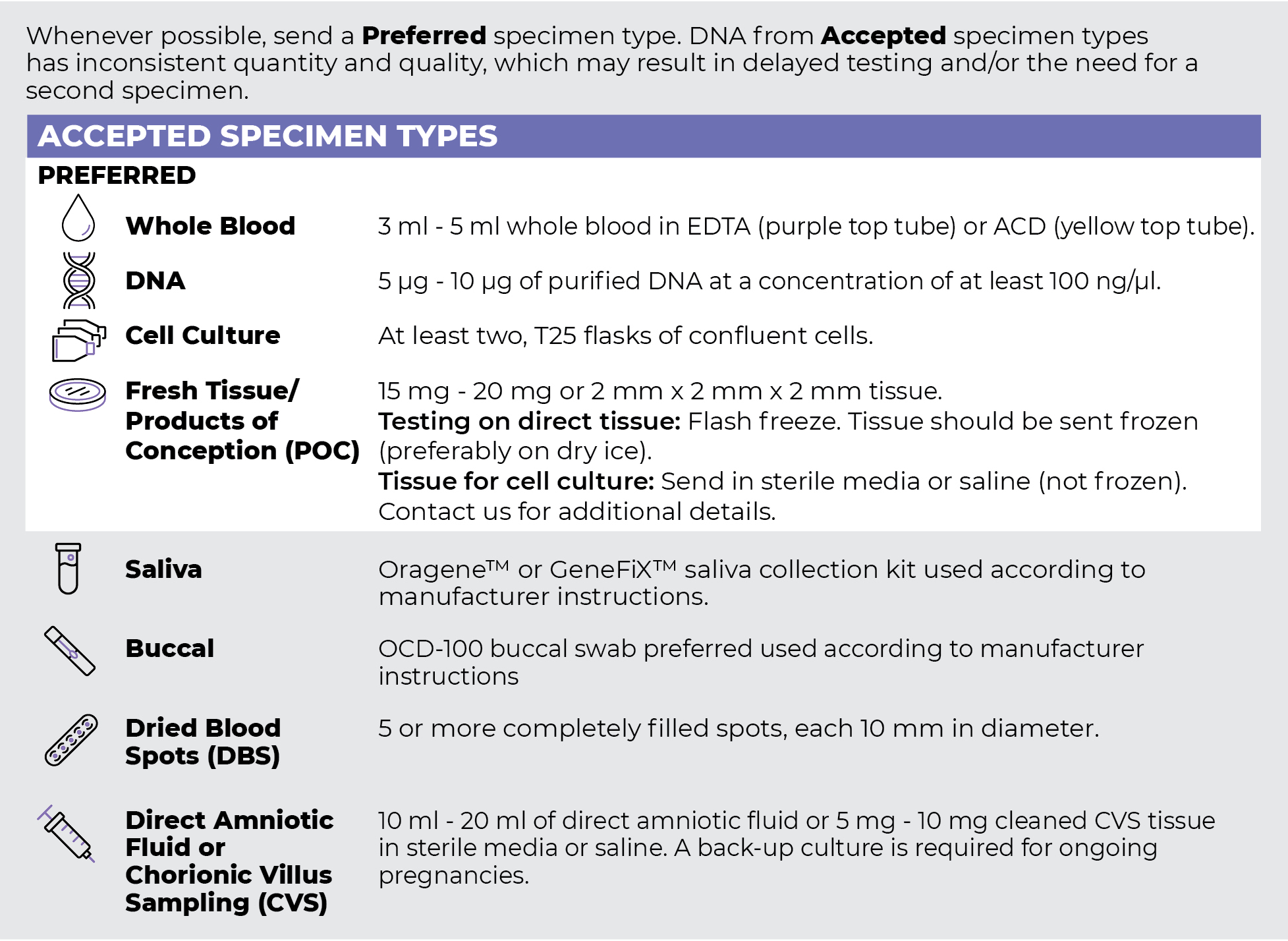
Specimen Types
Specimen Requirements and Shipping Details
PGxome (Exome) Sequencing Panel

PGnome (Genome) Sequencing Panel

ORDER OPTIONS
View Ordering Instructions1) Select Test Type
2) Select Additional Test Options
No Additional Test Options are available for this test.

